
The tissue images and MS data presented in this note were provided by Dr. Reid Groseclose and Dr. Steve Castellino from the Department of Bio-Imaging of GSK, 709 Swedeland Road, King of Prussia, PA 19406, USA.
Application & Background
Matrix-assisted laser desorption/ionization (MALDI) mass spectrometry (MS) imaging is a powerful analytical technique. It allows researchers to map the distribution of thousands of analytes in one run without labeling. High spatial resolution imaging analysis is especially useful in many applications, such as checking drug distribution in and across tissues, looking for biomarkers specific to tumor cells, or resolving biomolecules at the cellular level. To achieve high spatial resolution images, it is essential to evenly deposit matrix on tissue sections, without letting the matrix crystals grow too large. While some matrices are known to generate fine crystals, 2,5-dihydroxybenzoic acid (DHB), one of the most commonly used matrices, is known to form large needle-shaped crystals. The HTX TM Sprayer and HTX M5 Sprayer have many parameters that can be modified to optimize matrix deposition protocol, including flow rate, nozzle speed, number of passes, and nozzle temperature. In addition to these features, the HTX M5 Sprayer also features a heated tray that can precisely control the temperature of the sample slide while spraying MALDI matrix.
Experimental
Sample preparation
Rat brain embedded in 10% gelatin solution was fresh frozen and cryosectioned sagittally at a width of 12 microns using a Leica CM 3050 cryomicrotome. Tissues were thaw-mounted onto ITO coated glass slides and kept frozen at -80ºC until analysis. Prior to matrix deposition, slides were kept in a desiccator at room temperature for 30 minutes. No pre-treatment or washing step was used.
Matrix Application
Six different experimental conditions, detailed in Table 1, were tested prior to varying the tray temperature to optimize the other parameters for the matrix coating. For all conditions, DHB was applied to the slides at a concentration of 30 mg/mL (in 50:50 methanol:water) using the HTX M5 Sprayer.

Table 1 . Experimental conditions tested for optimal matrix coating.
Slides were then examined under the microscope for comparison (Figure 1). It was found that a matrix density of 8 μg/mm2, which corresponded to a nozzle velocity of 1000 mm/min (Conditions 1-4), gave good matrix coverage both on- and off-tissue. Matrix densities of 5.3 μg/mm2 (Condition 5) and 4 μg/mm2 (Condition 6) did not provide good coverage.
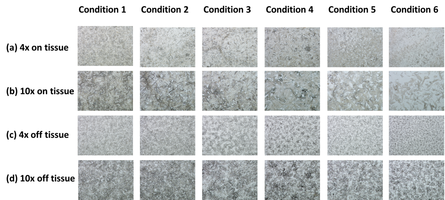
Figure 1. Microscopic pictures of matrix coated slides. (a) 4x magnified on-tissue images; (b) 10x magnified on-tissue images; (c) 4x magnified off-tissue images; (d) 10x magnified off-tissue images.
The final parameters of the HTX M5 Sprayer were thus set as displayed in Table 2 for the remaining MS imaging experiments. To minimize the time that the sample was exposed to an elevated temperature, all slides were kept off the tray until the target temperature was reached. The slides were then placed on the tray for 1 minute prior to spraying to allow the temperature to equilibrate. After spraying, the slide was taken off the tray and immediately placed in a desiccator.

Table 2. Spray parameters.
Mass spectrometry analysis
MS imaging was performed on slides prepared under experimental conditions 1-4 using a MALDI-Orbitrap system, where a MALDI/ESI injector (Spectroglyph) was coupled to an Orbitrap Velos mass spectrometer (Thermo Scientific) for high resolution MS analysis. The MALDI source was equipped with an Explorer One Nd:YLF (349 nm) laser firing at 500 Hz and 2.2 Amp. Mass spectra were acquired for m/z 150-2000, with a resolving power of 7500 (at m/z 400) and a maximum injection time of 50 ms (automatic gain control target = 1 x 10⁶). Rat cerebellum and brain stem areas were selected for imaging at a pixel size of 25 μm.
Results
When comparing slides with the same matrix density (Conditions 1-4), slides sprayed when the sample tray was at higher temperatures clearly displayed finer DHB crystals compared to ones sprayed when the sample tray was at lower temperatures. For example, 10x on-tissue microscopic pictures (Figure 1b) revealed fine crystal structures in Conditions 1 and 2 (55ºC and 45ºC, respectively), but visible needle-shaped crystal structures in Conditions 3 and 4 (35ºC and 25ºC, respectively). The same trend was observed in the off-tissue microscopic pictures. Therefore, we concluded that increasing the sample tray temperature during matrix spraying helped to reduce matrix crystal size, which increases the quality of high spatial resolution MS images.
A common lipid list of 350 species was used to map distribution, and selected ion images are shown in Figure 2. It was observed that the slides sprayed at higher temperature generated sharper, more intense, and more evenly distributed signals. On the other hand, similar number of peaks was detected across all 4 conditions (Figure 3), so there is no obvious evidence suggesting that elevated temperature causes degradation of lipid species. Further research is needed on the effect of elevated tray temperatures on MS imaging analyses when working with heat sensitive or labile analytes.

Figure 2. MS images of lipid species detected on rat brain sections at each condition. The m/z values, tentative lipid identification and MSI results are listed.

Figure 3. Spectra corresponding to a limited m/z range from each of the four experimental conditions tested.
Conclusions
Generating fine matrix crystals is essential for high resolution MS imaging experiments. Applying DHB matrix at elevated tray temperatures helped to create fine matrix crystals, which resulted in sharper and more intense MS image signals. Future research seeks to explore the effect of an elevated tray temperature on the quality of MS images across other analytes and matrices.